Eye Dissection Worksheet Answer Key
Eye Dissection Worksheet Answer Key - Find iris and remove it from eye. Examine the back of the eye and find extrinsic muscle bundles, fatty tissue and the optic nerve. Family nurse practitioner is a key resource for advanced practice nurses and graduate students seeking to test their skills in assessing, diagnosing, and managing cases in family and primary care. The lesson includes educational videos, an interactive quiz, a student checklist, an interactive laboratory powerpoint, and more!
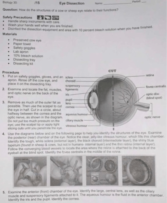
Solved Retina Biology 30 15 Eye Dissection Name Question
Protects the eye and helps it keep its shape. This lesson plan describes the cow eye dissection in detail. This cushions the eye from shock in its bony orbit.
Name The Three Layers That Make Up The Wall Of The Eyeball.
Outer layer of the eye. Identify the following external structures: Locate the covering over the front of the eye, the cornea.
It Contains Questions Asking Students To Observe And Describe The External Structures Of The Eye, Identify Internal Structures Like The Cornea, Iris, And Retina, And Their Functions.
Your studentsmore relevant and to reduce the number of specimens. Clean up and wipe down all lab dissection equipment. The sclera , the tough, external, white coat.
Study With Quizlet And Memorize Flashcards Containing Terms Like Anterior Chamber, Ciliary Muscle, Retina And More.
External anatomy look carefully at the preserved cow eye. Name the three layers that make up the wall of the eyeball. Start studying sheep eye dissection quiz.
In Your Cow's Eye, The Cornea May Be Cloudy.
Dry the eye with paper toweling. On the back of the eye, the thin layer of cells of the retina can be seen here, it is very thin and easy to pull away. Safety goggles • dissection scissors.
The Cow Eye Is Also Large Which Makes The Dissection And Identification Simple, But.
This document is a worksheet for dissecting and identifying the structures of a cow eye. Hole in the iris that allows light into the inner eye. Cord at the back of the eyeball.
You Should Be Able To Find The Sclera, Or The Whites Of The Eye.
Describe the function of the following structures: Cut away the fat and muscle. Make incision through sclera in the middle of the eye using scalpel.
Student Exploration Sheet, Answer Key, Teacher Guide, And Vocabulary Sheet Included.
External features of the eye. Colored ring of muscle that changes the size of the pupil. Outer lens that lets light into the eye.
Continuation Of The Optic Nerve That Relays Information From The Optic Chiasm.
The gelatinous fluid inside is the vitreous humor, the lens sits within this liquid. The cow eye is an excellent specimen to use, because it is very similar to the human eye which makes comparing the structures. Protects the eye and helps it keep its shape.
This Tough, Outer Covering Of The Eyeball Has Fat And Muscle Attached To It.
Learn how to dissect a cow's eye in your classroom. The white part of the eye, the sclera, is a tough, outer covering The conjunctiva , reflected over the anterior surface of the eye and attached to the eyeball a short distance from the edge of the.
Download Now And Let The Words Propel You.
A layer of cells in the back of the eye that picks up vibrations of visible. Using scissors, cut the eye in half. Perform a fully immersive virtual reality (vr) dissection of a human heart, cow eye, pig kidney, and sheep brain.
Cut Until Clear Liquid (Aqueous Humor) Is Released.
You may be able to look through the cornea and see the iris, the colored part of the eye, and the pupil, the dark oval in the middle of the iris. Remove cornea and cut it with scalpel. Learn vocabulary, terms, and more with flashcards, games, and other study tools.
The Coloured Portion Of The Eye.
Dissecting tray or plastic container. Locate the cornea, sclera, and optic nerve. Outer layer of the eye.
Use The Structures Listed In Question #2 And Label The Diagram.
Preserved cow's eye • dissection tray. The white part of the eye, the sclera, is a tough, outer covering of the. Separate the parts of the eye.
Name Three Structures That Help Focus The Light Rays Entering The Eye.
Sends messages from the eye to the brain. Name three structures that help focus the light rays entering the eye. When the cow was alive, the cornea was clear.
Cord At The Back Of The Eyeball.
Study with quizlet and memorize flashcards containing terms like optic nerve, lens, cornea and more. Locate cornea, sclera, and optic nerve. Cow eye dissection 2/6 cow eye observation:
Back Of The Eye (With Rods And Cones) (Rods See Shadows And Cones See Color) Location.
Wash the sheep eye in running water to remove the preservative fluid. Sign up and see the remaining cards. Forceps • vinyl or latex gloves.
In A Downloadable Pdf Format (*), This Ebook Is A Beacon Of Encouragement.
The clear gel that fills the space between the lens and the retina of the eyeball of humans and other vertebrate. When the cow was alive, the cornea was. Locate the cornea, sclera, and optic nerve.
Note The Fat (Adipose Tissue) On The Surface, Of The Eye.
The coloured portion of the eye. Family nurse practitioner is a key resource for advanced practice nurses and graduate students seeking to test their skills in assessing, diagnosing, and managing cases in family and primary care. Make incision into cornea with scalpel.
Use A Scalpel To Make An Incision In The Cornea.
Mammalian organ pack dissection by victoryxr: Examine the outside of the eye.

Eye Anatomy Worksheets Printable Worksheets

anatomy of the eye coloring worksheet answer key Anatomy Worksheets

Cow Eye Dissection Worksheet Answers prntbl.concejomunicipaldechinu.gov.co
Solved retina Biology 30 15 Eye Dissection Name Question

Frog dissection labeling worksheet Ojo Post Lab Questions The membrane holds the coils of the

The Language Of Anatomy Worksheets Answer Key Printable Worksheets

Lab5 Sheep eye dissection Part 1 Diagram Quizlet

Frog Dissection Worksheet Answer Key

SOLUTION Eye dissection Studypool

Solved COW EYE DISSECTION LAB SHEET Video Tutorial

Vision and the Structure of the Eye

43 cow eye dissection worksheet Worksheet Master

Eye Dissection

Dissection 101 Cow Eye Dissection Lesson Plan PBS LearningMedia

Practice Worksheets Parts Of The Eye Worksheet For 6t vrogue.co
